1. Executive Overview: What is a PTB Fracture Brace and Why is it Critical in Modern Orthopedics?
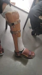
The Patellar Tendon Bearing (PTB) Fracture Brace represents one of the most significant advancements in non-operative and post-surgical lower-limb trauma rehabilitation. Designed under the classic biomechanical principles pioneered by Dr. Augusto Sarmiento, the PTB brace is an external rigid-shell orthosis specifically engineered to immobilize diaphyseal tibial fractures, distal tibial shaft fractures, and select delayed-union conditions while allowing controlled early weight-bearing.
For global healthcare buyers, orthopedic department heads, and rehabilitation clinic directors, procuring high-grade PTB Fracture Braces requires a clear understanding of the biomechanical mechanism: transferring axial ground reaction forces directly from the lower leg to the patellar tendon, anterior tibial flare, and medial femoral condyle. By bypassing the mid and distal tibia, a professionally fabricated PTB brace reduces total axial load on the fracture site by 30% to 50%, depending on patient anatomy and shell contouring.
Clinical Insight: Hydrostatic Fluid Dynamics & Bone Healing
Modern functional bracing does not rely solely on mechanical bone rigidness. The enclosed PPCP (Polypropylene Copolymer) shell generates a closed hydrostatic chamber within the soft tissue compartments of the calf. As the patient muscles contract against the rigid shell wall during ambulation, fluid pressure rises, distributing axial forces uniformly around the limb. This micro-motion at the fracture site actively stimulates osteogenesis and accelerates secondary callus formation without inducing angular deformity.
Biomechanical Load-Sharing Breakdown of the PTB System
To evaluate the functional efficacy of a PTB Fracture Brace during clinical tender evaluations, purchasing committees must examine the four primary anatomical load-bearing surfaces:
- Patellar Tendon Indentation (Patellar Bar): Positioned at the mid-point of the patellar ligament (between the lower pole of the patella and the tibial tuberosity). This deep indentation acts as the primary vertical weight-bearing shelf.
- Anterior Tibial Flare Contour: Spreads load across the broad, non-tender medial flare of the proximal tibia, preventing localized pressure necrosis.
- Posterior Popliteal Wall Pressure: Provides an opposing anteroposterior (A-P) force vector that locks the patellar tendon into its designated shelf during knee flexion up to 90 degrees.
- Medial & Lateral Condylar Wedges: Encase the femoral condyles to prevent frontal-plane (valgus/varus) displacement during stance phase.